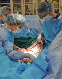
CALEC surgery, or cultivated autologous limbal epithelial cell surgery, represents a groundbreaking advancement in eye treatment, particularly for patients with severe corneal damage. This innovative procedure utilizes stem cell therapy, allowing medical professionals to harvest limbal epithelial cells from a healthy eye and cultivate them into a cellular graft that can regenerate the damaged cornea. The promising results from clinical trials demonstrate that this approach not only restores the cornea’s surface but also offers hope to individuals previously deemed unsuitable for corneal transplants. With over 90 percent effectiveness, CALEC surgery showcases the potential of regenerative medicine in cornea restoration, addressing the urgent need for effective solutions in treating debilitating corneal injuries. Such advancements not only enhance vision recovery but also significantly improve the quality of life for patients suffering from vision-threatening conditions.
In recent years, the field of ocular medicine has witnessed remarkable progress, especially with techniques involving limbal cell transplantation. The utilization of autologous stem cell grafts to rejuvenate the corneal surface offers a promising alternative to traditional corneal transplant methods. By leveraging the body’s own limbal epithelial cells, eye specialists are redefining standards for treating blinding corneal injuries. Innovative approaches, such as CALEC surgery, highlight the transformative potential of regenerative therapies for restoring sight and revitalizing patients with previously untreatable corneal damage. This method not only exemplifies cutting-edge scientific advancements but also underscores the growing importance of personalized healthcare solutions in ophthalmology.
Innovative Approaches in Eye Treatment: The Role of CALEC Surgery
The groundbreaking CALEC surgery represents a significant advancement in surgical techniques aimed at restoring vision to patients with corneal damage. By employing cultivated autologous limbal epithelial cells, this innovative method enables the transplantation of healthy stem cells from the patient’s own eye to repair the damaged cornea. As a result of careful research and clinical trials, CALEC surgery has demonstrated over 90% efficacy in reinstating the corneal surface, offering new hope to individuals who previously faced irreversible vision loss due to severe corneal injuries. This pioneering approach could revolutionize eye treatment methodologies comparing favorably to traditional corneal transplants, emphasizing the need for further studies to enhance its application across a broader patient base.
The implications of successful CALEC surgery extend beyond the immediate restoration of vision. With the ability to regenerate vital limbal epithelial cells, this surgery not only aids in the immediate healing of the cornea but also significantly reduces the chronic pain and visual complications that plague patients suffering from limbal stem cell deficiency. As the CALEC method advances, researchers are optimistic about refining techniques, particularly by exploring the possibility of allogeneic grafting, which would allow for broader applications, including in patients with bilateral eye damage. Such developments may redefine standards in eye treatment, providing a novel option for those long considered untreatable.
Frequently Asked Questions
What is CALEC surgery and how does it use stem cell therapy for cornea restoration?
CALEC surgery, or cultivated autologous limbal epithelial cells surgery, involves extracting limbal epithelial cells from a healthy eye, expanding them into a graft, and transplanting this graft into a damaged cornea. This innovative eye treatment utilizes stem cell therapy to restore the cornea’s surface, providing hope for patients with blinding corneal injuries that were previously considered untreatable.
What are the main benefits of CALEC surgery compared to traditional corneal transplant procedures?
CALEC surgery offers several advantages over traditional corneal transplant methods, including a more than 90% success rate in restoring corneal surfaces and significantly reduced risks of transplant rejection. By using stem cells from the patient’s own healthy eye, this treatment enhances the potential for better integration and healing, making it a promising alternative for individuals with limbal stem cell deficiencies.
How effective is CALEC surgery in treating corneal surface damage?
Results from clinical trials indicate that CALEC surgery is highly effective, with complete restoration of the cornea reported in 50% of participants by three months, rising to 79% at 12 months. The overall success rates were 93% and 92% at 12 and 18 months, respectively, showcasing its significant potential to improve vision in patients with serious corneal damage.
What are limbal epithelial cells, and why are they important in CALEC surgery?
Limbal epithelial cells are specialized stem cells located at the eye’s limbus, crucial for maintaining the cornea’s smooth surface. In CALEC surgery, these cells are harvested from a healthy eye, expanded, and used to repair a damaged cornea. Their role is vital, as they help restore normal corneal function and surface integrity in patients with severe corneal injuries.
Is CALEC surgery available as a standard treatment at hospitals?
Currently, CALEC surgery remains experimental and is not offered as a standard treatment in hospitals, including Mass Eye and Ear. Ongoing studies are necessary to gather more data before seeking federal approval. Researchers hope that successful trials will lead to wider availability of this revolutionary eye treatment in the future.
What are the potential risks associated with CALEC surgery?
The clinical trials for CALEC surgery have demonstrated a high safety profile, with no serious events reported in donor or recipient eyes. However, minor adverse reactions, such as a bacterial infection due to chronic contact lens use, have occurred. As with any surgical procedure, discussions regarding risks and benefits are essential before undergoing treatment.
| Key Points |
|---|
| Ula Jurkunas performs first CALEC surgery at Mass Eye and Ear, highlighting innovative procedures for eye damage. |
| CALEC surgery involves harvesting stem cells from a healthy eye and transplanting them into a damaged eye, aiming to restore corneal surfaces. |
| The clinical trial achieved over 90% effectiveness in restoring corneal surfaces in patients after 18 months of follow-up. |
| Patients experience relief from pain and visual difficulties caused by limbal stem cell deficiency. |
| The CALEC procedure is currently experimental and not available in U.S. hospitals; additional studies are needed for federal approval. |
| Future research aims at expanding treatment availability possibly using donor stem cells from cadaveric eyes. |
| The National Eye Institute funded this innovative study, marking it as the first human stem cell therapy in the U.S. |
| A collaborative effort among prestigious institutions like Dana-Farber and Boston Children’s Hospital enhanced the success of this clinical trial. |
Summary
CALEC surgery represents a groundbreaking advancement in corneal treatment, providing new hope for patients with severe eye injuries previously deemed untreatable. This innovative approach utilizes stem cells harvested from a healthy eye to effectively restore corneal surfaces, demonstrating over 90% effectiveness in clinical trials. As the research progresses, the potential for broader applications is on the horizon, paving the way for revolutionary changes in ocular therapeutics.

